Wellcome to the website of scientific journal «Morphological Newsletter - Morfologicheskie Vedomosti»!

"Morfologicheskie Vedomosti - Morphological Newsletter" is the Russian national biological scientific quarterly journal, publishes the latest achievements in the field of sciences about the development, morphology and structure of the animals and human body including experimental morphology. The priorities of the journal are scientific achievements and researches in the field of experimental and clinical anatomy, physical anthropology, animals and human development biology, neurobiology, cell biology, morphomics, pathology and of their teaching problems.
The journal is indexed in over than 30 world scientometric and information databases, including CrossRef, International Committee of Medical Journal Editors (ICMJE), World Association of Medical Editors (WAME) and is received to main Russian national libraries, National Library of Medicine (NLM) of USA, German Electronic Journal Library (EZB), China Knowledge Resource Integrated Database (CNKI).
Field of research results, published in the journal corresponds to the ciphers of the international UDC 611 - anatomy and comparative anatomy, 572.5 – somatology, 572.7 - morphology
Current issue
ONLINE ISSUE COVER (ISSN 2686-8741)
ONLINE TITLE PAGES (ISSN 2686-8741)
RESEARCH ARTICLES
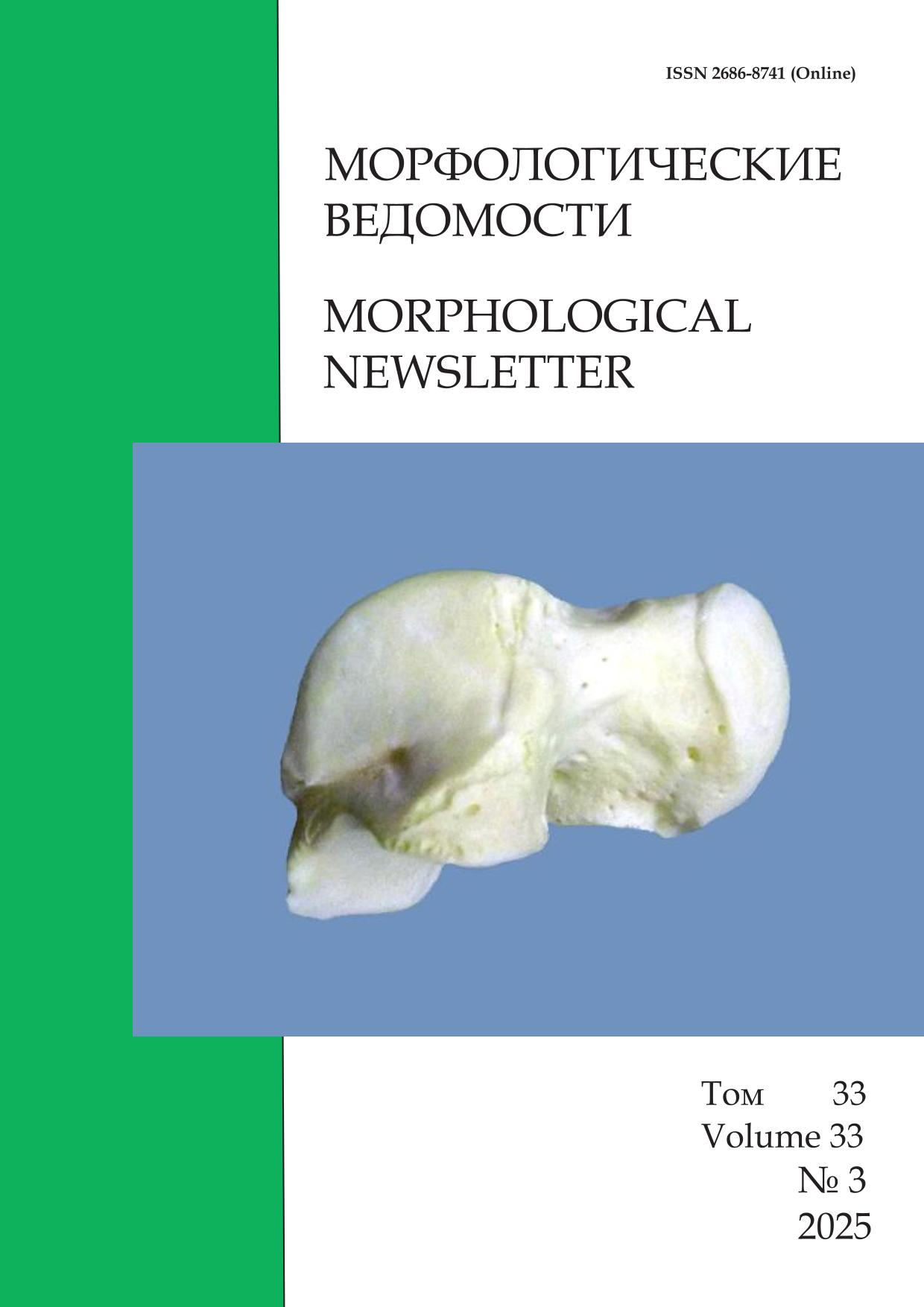
The author established anatomical variability of osteometric parameters of the talus in the Russian population and identified significant gender differences in them
The relevance of studying the talus anatomical variability is determined by the need to improve the accuracy of anthropological research in the context of diagnosing the ethnological and archaeological identity of finds. Its anatomical variability can serve as an important indicator of the social and cultural structure of modern and ancient populations and can also provide insight into the impact of environmental factors and lifestyle. The aim of this study was to establish the variability of osteometry parameters of the talus in adults using the Russian population as an example. The study included 75 talus specimens obtained from certified bone collections, which included 49 male and 26 female bones from cases aged 20 to 90 years. Seventeen osteometry parameters were measured, describing the linear characteristics of the talus, as well as its weight, volume, and density. Analysis of these variability parameters demonstrated significant sex differences in the osteometry and physical characteristics of the talus. In men, the greatest variability was observed in the parameters of neck and head length, posterior trochlea width and inferior articular surface, while in women - in the length of the neck and head and posterior width indicators. In both groups, low variability is characteristic of the height indicators of the anterior and posterior height. The most variable parameters of male bones are the width and length of the neck and head with a variation coefficient of 11.99% and 12.8%, respectively, and the least variable were the anterior height (4.53%) and posterior height (4.73%). The most variable parameters for females are the length of the neck and head and the width of the head with a variation coefficient of 12.7% and 12.4%, respectively, and the least variable, as in male talus bones, were the anterior height (4.26%) and posterior height (5.12%). The obtained data on the variability of osteometry parameters of the talus should be taken into account in anthropological and archaeological studies, clinical practice when planning surgical interventions and developing optimal methods for diagnosing and treating its pathology.
SHORT ARTICLES
It has been experimentally proven that bilateral ovariectomy causes uterine tubes epithelium morphological and functional changes
Ovariectomy leads to hormonal dysfunction, which manifests itself at all levels of the structural organization of the female reproductive system, including the epithelial lining of the uterine tubes. The aim of this study was to investigate the morphological and functional characteristics of the epithelial lining of all sections of the uterine tubes in reproductive-aged rats with bilateral ovariectomy. Twenty-six-month-old female Wistar rats served as subjects for the morphological study. All animals were divided into two groups: 10 females in the control group and 10 females with ovariectomy. Ovariectomy was used as an experimental model to exclude the influence of ovarian hormones on the uterine tubes. The uterine tubes were fixed in 10% buffered formalin, then paraffin sections (4 μm thick) were cut and stained with hematoxylin and eosin. Cell counts were calculated based on at least 1000 cells per animal. Morphometric analysis of tissue components of the epithelial lining of the uterine tube mucosa was measured using the ImageJ software. Estrous cycle phase was determined by staining vaginal smears using the RAPIHEM protocol. Rats in the control and experimental groups were in the resting phase. Fisher's exact test was used to test for homogeneity of variances. Differences were considered statistically significant at a significance level of p < 0.05. After bilateral ovariectomy, the uterine tube epithelium undergoes significant restructuring in all sections, manifested by an increase in mucosal thickness and a transformation of the cellular composition. The greatest changes are observed in the ampullary and uterine sections of the uterine tubes. The ampullary section is characterized by a 2.6-fold decrease in intercalated cells and a 3.5-fold increase in secretory cells. For the uterine section, the percentage of secretory cells increases by 2.0. The ciliary beat frequency of the uterine tube epithelium decreased by 40% in all regions compared to the control group. These changes are a potential risk factor for uterine tube dysfunction.
The results of the study show that experimental masticatory hypofunction causes progressive atrophy of the alveolar bone tissue, degeneration of the periodontal ligament, activation of osteoclasts, and a decrease in the resistance of the periodontium to mechanical load
The studying the influence of masticatory load on the structure and function of the periodontium is an important area of research in dentistry. Disruption of occlusal relationships, the absence of teeth, or prolonged use of a soft diet led to hypofunction of the masticatory apparatus, which provokes atrophic and dystrophic processes in periodontal tissues. Masticatory load is a key factor maintaining periodontal homeostasis. The aim of this study was to establish patterns of structural and functional reorganization of the periodontium during experimental masticatory hypofunction. The temporal and qualitative characteristics of morpho-functional changes in the periodontium were studied during experimental modeling of masticatory hypofunction. Sixty mature outbred white rats (6 months old, weighing 250-300 g) were used in the study. The animals were divided into 2 groups. The main group (n=45) with experimentally modeled masticatory hypofunction, and the control group (n=15) consisted of intact animals. Masticatory hypofunction was simulated by feeding the animals soft food dispersed to a puree state. The experiment lasted 12 weeks. The results were evaluated using a micro-computerized X-ray method, histological, immunohistochemical, molecular biological, and biomechanical analyses. After 12 weeks of hypofunction, the bone volume to total bone tissue ratio decreased by 29% (<0.001), indicating pronounced bone resorption. The trabecular thickness decreased by 33%, and the number of trabeculae decreased by 34% (<0.001), which generally confirms a disruption of bone tissue microarchitecture. Histological studies showed that by week 12 of the experiment, the periodontal ligament thickness decreased by 38.7%, the osteocyte count decreased by 42.3%, and the osteoclast count increased by 2.1 times. Molecular biological changes relative to the control included a 0.6-fold decrease in the synthesis of the alpha chain of type I collagen and a 0.5-fold decrease in alkaline phosphatase activity, as well as a 1.8-fold increase in the activity of tartrate-resistant acid phosphatase (all changes are significant (p<0.001). The results of the experimental study explain the accelerated tooth loss in patients with unilateral chewing.
It has been established that during physiological pregnancy, the morphological and functional parameters of the umbilical cord and its vessels retain their structure during labor with an initially normal anatomical structure
A number of umbilical cord morphometric parameters, including its circumference, umbilical vessel diameters, umbilical cord area, umbilical arteries and veins, and embryonic mucosal connective tissue, are not subject to mandatory ultrasound examination. However, assessing these morphometric parameters during the antenatal period could help predict the outcome of labor. The aim of the study was to evaluate the impact of labor on the morphological and functional parameters of fetal umbilical vessels during full-term pregnancy. A total of 150 pregnant women participated in the study. The examination was conducted using a Samsung Accuvix XG 2019 ultrasound machine during the active phase of labor during contractions and 2-3 days before the onset of labor. The following anatomical and functional parameters were recorded: umbilical cord circumference in the proximal, median, and distal segments; internal diameters of the right and left umbilical arteries and vein in three similar segments; pulsatility index; maximum systolic blood flow velocity; and end-diastolic blood flow velocity. The cross-sectional areas of the umbilical cord, arteries, veins, and embryonic mucosal connective tissue were calculated. Statistically significant differences were found in maximum systolic and end-diastolic blood flow velocities, and consequently, in the pulsatility index. According to the nonparametric Wilcoxon test, umbilical cord circumference, internal diameters of umbilical vessels, and cross-sectional areas undergo statistically insignificant changes. The volume of embryonic mucosal connective tissue has a greater influence on umbilical cord circumference and cross-sectional area. With a normal umbilical cord anatomical structure, the intrapartum period proceeds without complications, despite fluctuations in hemodynamic parameters. Morphological parameters, namely, umbilical cord circumference in the proximal, medial, and distal segments; the internal diameter of the right and left umbilical arteries and vein in three analogous segments; the cross-sectional areas of the umbilical cord, arteries, and vein; and the embryonic connective tissue mucosa, retain their structure during the intrapartum period despite initially normal anatomical structure. The obtained results can be used as predictors for assessing birth outcomes.
SCIENTIFIC REVIEWS
A systematic literature review of the hyaluronic acid-enriched culture medium use in embryo transfer to improve outcomes in assisted reproductive technology programs
Infertility is a global problem. According to the data of the World Health Organization, 17.5% of people, or one in six people worldwide, suffer from infertility as of 2023. Experts believe that by 2050, a critical decline in the birth rate will lead to a reduction in the number of children under 15 by 40% or more. In Russia, statistics reflect global trends. Therefore, the search for new, effective infertility treatments is a pressing issue in modern medicine. The objective of this review was to analyze and evaluate current data on the use of a hyaluronic acid-enriched medium during embryo transfer to increase implantation rates in infertility treatment cycles using assisted reproductive technologies. The sources of specialized literature used in this review were electronic libraries of scientific publications and medical databases such as Google Scholar, PubMed, and elibrary.ru. The review included relevant publications found through a search using the following keywords: "assisted reproductive technologies," "hyaluronic acid," "embryo," and "endometrium." The study found that the Russian Federation ranks first among European countries and fourth globally in the number of assisted reproductive technology cycles performed (140,931 cycles in 2020 and 158,893 in 2021). However, according to the Russian Association of Human Reproduction and the European Society of Human Reproduction and Embryology, the effectiveness of assisted reproductive technology programs is no more than 40%. Infertility treatment with assisted reproductive technology can be unsuccessful for a number of key factors, including the lack of effective embryo implantation in the endometrium. One of the leading causes of implantation failure of a competent embryo is the failure to form an adhesive matrix between the embryo and the endometrium. The coordinated differentiation of endometrial cells and their interaction with the embryo play a crucial role in this process. It is hypothesized that the use of hyaluronic acid-enriched culture media during embryo transfer may help address the problem of implantation failure in assisted reproductive technology programs. A systematic review of the literature on the use of hyaluronic acid-enriched culture media during embryo transfer to improve assisted reproductive technology outcomes was conducted. Possible mechanisms of action for this medium during assisted reproductive technology embryo transfer are described.
HISTORY OF MORPHOLOGY
The article is dedicated to the 160th anniversary of the birth of the famous Russian Moscow anatomist, Professor Peter Karuzin
Peter Karuzin (1864–1939) is one of the significant figures in the history of Russian anatomy and medical science during the first half of the twentieth century. Born in St. Petersburg, he developed an early interest in studying human body structure from childhood. He received his education initially at a gymnasium before enrolling in the Medical Faculty of Moscow University where he distinguished himself as a diligent student and promising researcher. After graduating from university in 1889, he began his scientific and pedagogical career on the department of normal anatomy under the leadership of Professor Dmitry Zernov. It was under this professor's guidance that he defended his doctoral dissertation, which became the first major Russian research dedicated to spinal cord morphology. This work marked the beginning of his active academic and teaching career. In subsequent decades, Peter Karuzin combined scientific activity with intensive teaching responsibilities. He supervised practical sessions and developed new teaching methods by successfully incorporating modern European approaches into anatomy instruction. His trip abroad towards the end of the nineteenth century played a particularly important role, as it introduced him to European traditions of anatomical research and educational practices, enriching his knowledge base and serving as a foundation for reforming educational practice in Russia. One of Peter Karuzin's greatest achievements was designing and constructing the modern Anatomy Building of Moscow University. Built between 1926 and 1928, this unique facility provided optimal conditions for training future physicians and conducting scientific research. For many years, Peter Karuzin personally taught numerous courses and conducted practical lessons, preparing a new generation of qualified specialists. Beyond teaching, Peter Karuzin carried out important studies focused on brain and spinal cord structures and functions, developing principles for differentiated study of neural pathways. Additionally, he authored a foundational guidebook on plastic anatomy, widely used among artists and medical professionals alike. The legacy of Peter Karuzin spans a broad range of areas, encompassing both theoretical foundations and methodological advancements in the field of anatomy, as well as direct influence over the formation of generations of Russian medical scientists.
ISSN 2686-8741 (Online)


